| 今月の症例 | 資料室 | ニュース | メールサービス | 憩いの部屋 | 歯科リンク
& Top |
| 今月の症例 | 資料室 | ニュース | メールサービス | 憩いの部屋 | 歯科リンク
& Top |
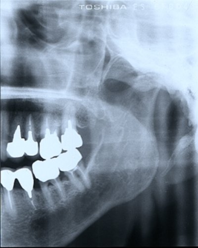
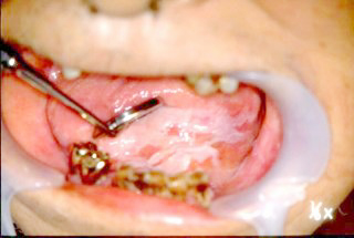
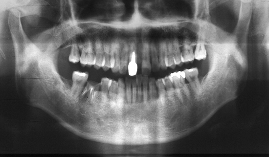
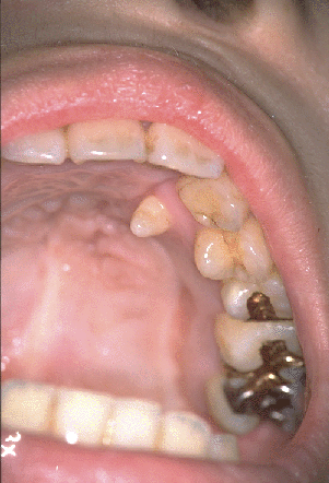
Horizontally Impacted third molar.
 Fig.
1 at the age of 16. Fig.
1 at the age of 16. |
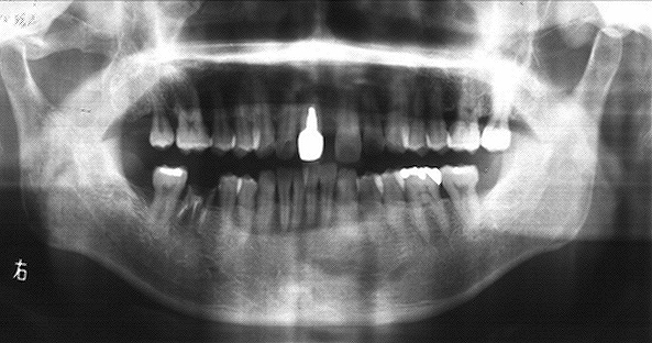
 Fig.
2 at the age of 23. Fig.
2 at the age of 23. |
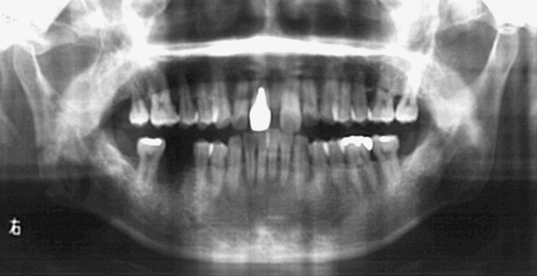
 Fig.
3 at the age of 27. Fig.
3 at the age of 27. |
**** < 今月の症例 > ****
2002年4月
lipoma 2002/4/3
| 脂肪腫:
Lipoma; 口唇粘膜に広基底性、弾性軟の
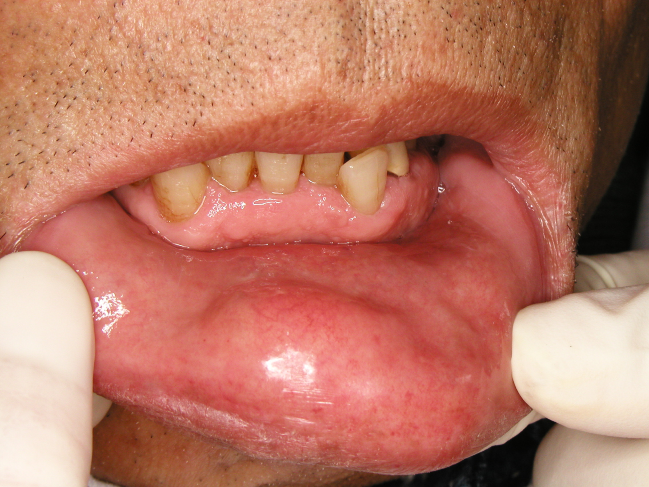
Patient. A 62-year-old man presented complaining of a "swelling" in his labial mucosa of lower lip. History. The patient had noticed a small lumps in his lower lip ca. 10 years previously. The tumor had increased in size gradually, resultant deformity of lower lip had become a problem. Oral examination. A multi-lobulated, non-movable, firm nodulous mass about 2.5*3cm. in diameter was palpated in labial mucosa of the lower lip. Physical examination. This was essentially negative. Tentative diagnosis. Lipoma. Operative procedure. Under local anesthesia a 3 cm. horizontal incision was made, with a No. 15 blade, in the mucosa of the lip. The incision was held open with Allis forceps and the specimen was " shelled out" and cut from its only attachment to the lip muscle by scissors, and removed. The incision was closed with 000 nylon suture. Microscopic Diagnosis. Lipoma. |
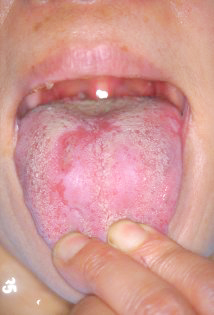
口唇炎;舌炎;甲状腺腫:Cheilitis, Glossitis & Goiter: |
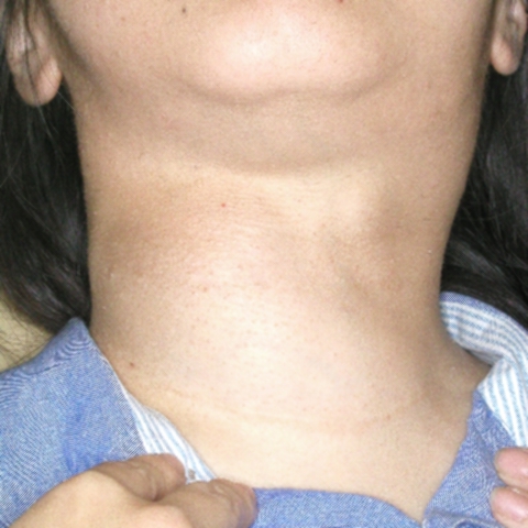 Goiter Goiter
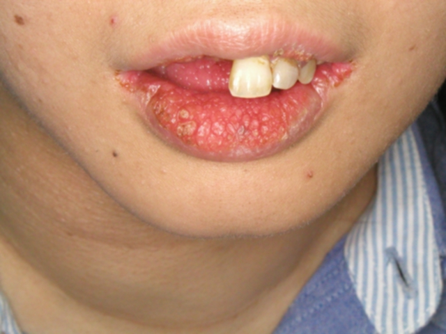 Cheilitis Cheilitis
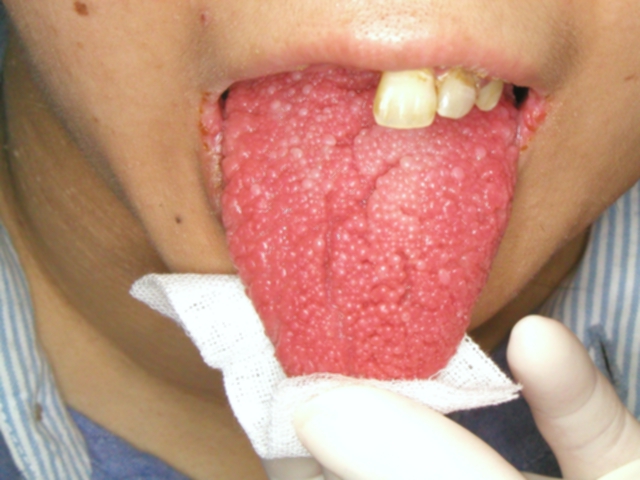 Glossitis Glossitis |
|
A patient, 27-year-old female, was referred to us for multiple lymph node swelling of the neck and submandibular region. Pt. had had operated on the thyroid 10 years ago. Concerning the tumor Pt. was referred to the specialist. Interestingly a cheilitis and a glossitis were
(Actinic) Cheilitis:
Glossitis
27歳、女性、顎下部および頸部の多発性リンパ節腫脹の精査目的に当科を紹介されたものですが、甲状腺、リンパ節腫脹等に関しては10数年前に手術を施行した施設に紹介、転院しまた。
|
Congenital ankyloglossia and frenectmy
|
2001年12月
Dislocation of the mandible
片側性顎関節脱臼:Unilateral Dislocation of the Mandible: |
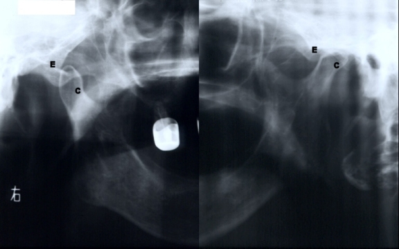
Radiographic Examination:Panoramic radiographs of the temporomandibular joint revealed the crest of the right condyle(c) anterior to and superior to the crest of the right articular eminence(e), while the left condylar head was in the glenoid fossa. |
| Patient:A 81-year-old woman was came to our OPD on November
11, with a dislocation of her mandible that had been present for 7 days. Treatment: To attempt manual reduction of the dislocation was successful. 81歳、女性、7日続く閉口不能による咀嚼障害で当科受診。 徒手整復を施術 |
2001年12月
リガ・フェーデ病:Riga Fede's disease生後34日、授乳障害の為来院 |
|
|
| Pt, 34-days-old baby, was brought to
our OPD for difficulties during breast feeding, traumatic ulceration on tongue/frenum. Treatment was extraction of the incisors. |
2000年12月
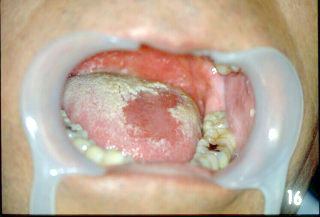
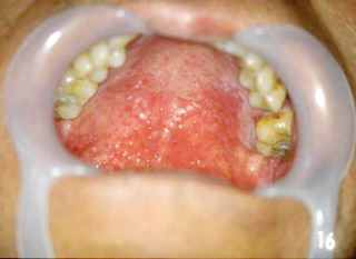
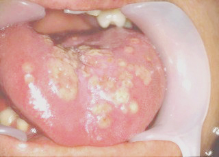
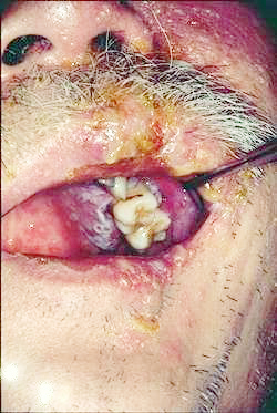
| ベーチェット症候群;再発性潰瘍
Behcet's syndrome; recurrent aphthous ulser (RAU)
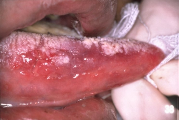
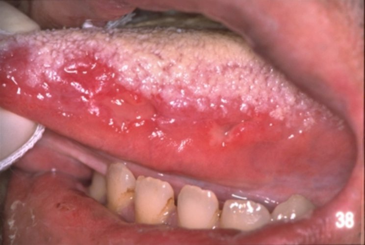
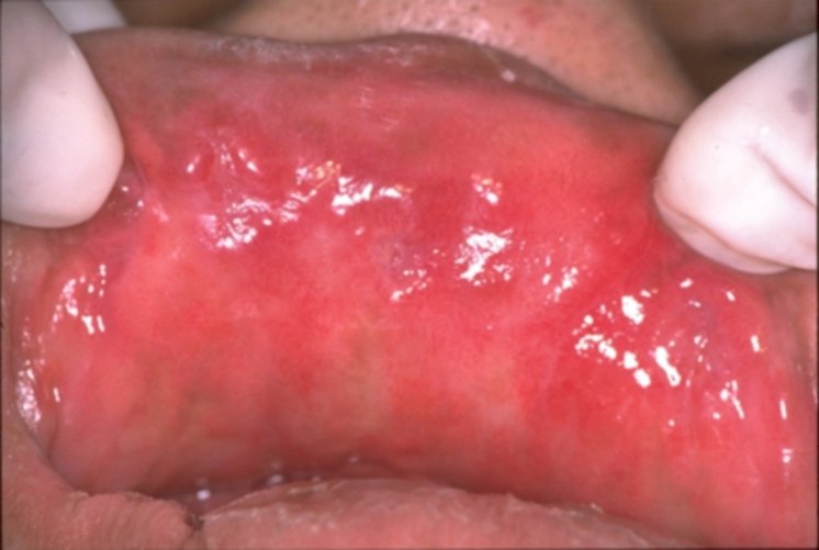
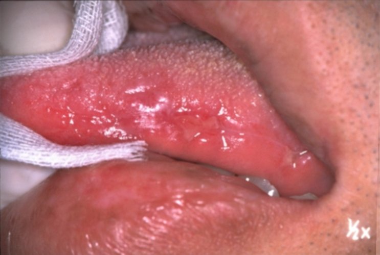
1998年2月に初発, 2000年7月急性化の為入院(3回目) 主訴:口内痛の為摂食不能 著明な舌の白苔, 小潰瘍を舌, 口唇, 頬粘膜に認める (Fig..1,2,3-5) ファンギゾンを投与,5日後白苔,疼痛は寛解し摂食可となる。 残存する粘膜潰瘍(Fig. 4,5-5) *口腔粘膜のアフタ性潰瘍・外陰部潰瘍・大腿部毛嚢炎・
写真をクリックしてご覧下さい
Male, 31 years, had a typical case of Behcet’s disease of 2 years duration. Note white coated tongue with apthous ulcer on the tongue at the first examination (Fig. 1,2,3-5). He was started on Fungisone. In a few days the pain and coated tongue was subsided (Fig. 4,5-5). |
|||||
| remark:
Common manifestations (over 90 % of patients) Recurrent oral ulceration (+) Genital ulceration (+) Eye lesions(uveitis) Less common manifestations Skin rashes and pustules (+) Arthritis of major joints (+) Neurological lesions ----- diffuse and intacranial Haematuria Abdominal discomfort (+) |
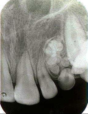
左側上顎乳犬歯の直上に歯牙様塊状物が認められる。犬歯は側方に転位し未萌出の状態である。
|
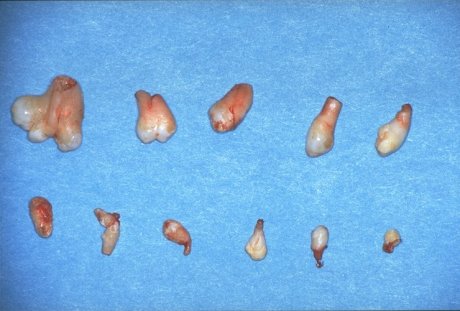
摘出された歯牙様硬固物
Tooth forms, single and fused, that were removed.
|
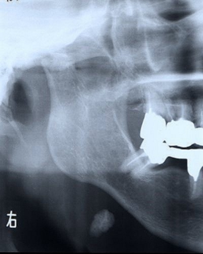
2000年10月
| 含歯性 ( 濾胞性 ) 嚢胞
Dentigerous (follicular) Cyst 当科を紹介され来院.上顎正中部に過剰歯と鼻腔底, 上顎洞に接するX線透過像を認める. 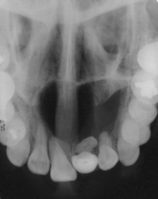
|
2,000年9月
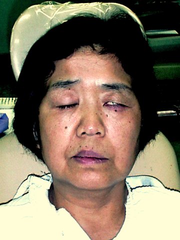
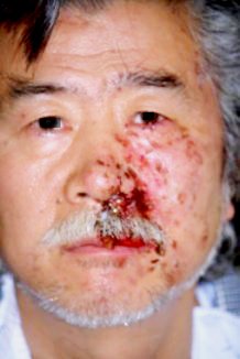
| 顔面神経麻痺
Bell’s palsy-----Facial paralysis
(意識的眼裂閉鎖不能) 口笛不能,鼻唇溝の消失,前額部皺の消失が認められる。
Female, 59 years, had a typical case of Bell’s palsy of 3 days duration. Note inability to close the upper lid and turning up of the eyeball (Bell’s sign ) and wrinkle of the forehead. Paralysis of the left side of the lip thus whistling or smiling is not possible. She was started on Predonosolone, 30 mg, two times a day. In 12 days the paralysis was markedly improved.
|
症例 8: 84歳女性 本院内科にて加療中の患者さんです。
両側顎関節(関節頭)の著明な変形(平坦化)が認められます。
尚右側顎下腺に唾石が認められます。
自覚症状は何れにもありません。
Case 8: A patient is a 84 year old female
who has been treated
for rheumatoid arthritis by an internist.
Panoramic radiograph showing bilateral
degenerative joint disease.
Bilateral flattening is seen in the temporal
components as well as in the condyles.
慢性関節リウマチは自己免疫不全状態を背景に持つ慢性の全身性炎症性疾患です。
その主病変は末梢の関節に発症します。顎関節の発症は少なからず診られるとされるのですが、歯科で診ることはやはり希です。
主な病態は滑膜の肉芽腫性病変です。増殖した滑膜は軟骨、軟骨下骨へと浸潤し、関節の破壊を来し関節の変形や運動障害を起こします。
治療方法は抗リウマチ剤-DMARD-等ありますが効果は不確実で原因療法は確立されていません。
急性期では、非ステロイド系抗炎症剤の投与と顎関節の安静、軟らかい食事の摂取、微温湯による湿布などの対症療法です。重症例ではhydroxychloroquine,
gold, penicillamine等で疼痛と炎症を制御します。
症状の寛解が認められたら、顎の可動性を確保するために軽度の顎運動を開始します。
顎関節強直症を後遺した時は病勢の停止を待って手術の適応と成ります(顎関節受動術)。
2,000年7月
耳下腺管憩室
Diverticulum of parotid duct
No. 9: 患者さんは、数十年余、左側頬部の無痛性腫脹を自覚しておりました。
起床時は、無症状ですが時間と共に漸次頬部は腫脹してくるとの由、
皮膚側より圧迫しますと、”ジュウ”といった感じが口内に走り腫脹は消失
していたようです。
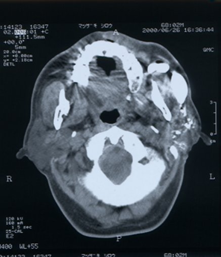
*Legend: Male, 68 years. He's noticed a chronic painless swelling
on the l-buccal region for decades of life. He has applied a pressure over
it to get relief. CT sialograms revealed Stenon's duct had two large diverticula.
2000年5月
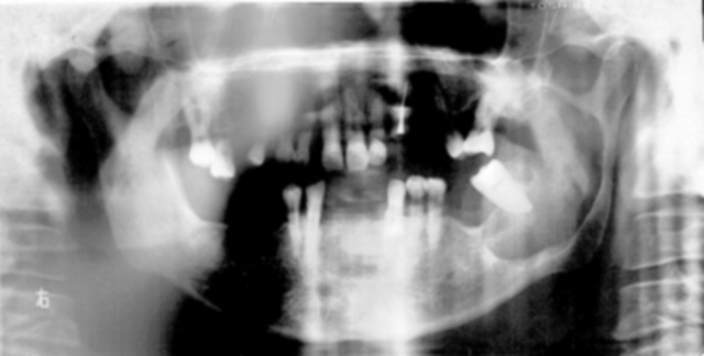
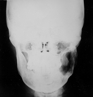
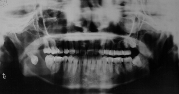
今週の症例はエナメル上皮腫と骨髄炎のX線写真と
歯牙の異所萌出です。
*註
エナメル上皮腫1-3、2-3:左側下顎遇角部より上行枝にわたる症例、開窓し経過観察中
エナメル上皮腫3-3:右側下顎骨体部の症例、開窓法は無効で、切除術の適応例
急性下顎骨骨髄炎1-4:初診、原因歯6の根尖に限局した透過像を認める他所見無し
急性下顎骨骨髄炎2-4:PCを7日投与(4g/日)間欠性疼痛は持続、CLDMに変更し奏功を得る
切痕部より後縁に斜走する透過像と遇角部に斑状透過像
急性下顎骨骨髄炎3-4:通院加療中(CLDM4週間)切痕、遇角部の皮質を含めた骨溶解像が明瞭
急性下顎骨骨髄炎4-4:通院加療中(CLDM6週間)X線透過像は若干不明瞭となる
*Legend
ameloblastoma 1-3: a large radiolucent lesion of the left side of mandible,
fenestrated
ameloblastoma 2-3: ib. 1-3, Posteroanterior view demonstrating large,
multilocular,
expansile radiolucent area.
ameloblastoma 3-3: circumscribed radiolucent area containing mandibular
right third molars.
fenestrated but failed to response.
acute osteomyelitis 1-4: Panoramic radiograph showing no radiolucent
lesion of the mandible.
acute osteomyelitis 2-4:CLDM relieved symptoms and X-P demonstrated
radiolucent lesion
around incisula and angular region. (treatment with CLDM, 2 weeks long)
acute osteomyelitis 3-4: radiolucent lesion getting obvious (treatment
with CLDM , 4 weeks long)
acute osteomyelitis 4-4: radiolucent lesion getting narrower
(treatment with CLDM, 6 weeks long)
Home|Previous Page|Next Page
1999年12月
悪性血管外(周)皮腫
malignant hemangiopericytoma
|
Fig. 8-1 |
Fig.8-2 |
Fig.8-3 |
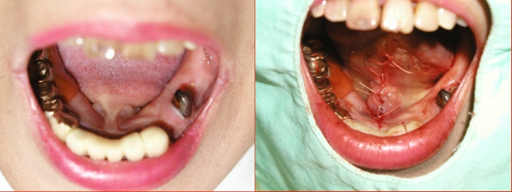
左側口蓋の血管外(周)皮腫
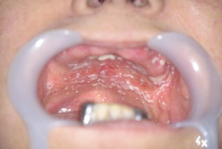
1998/7/20 患者さんは21歳、女性
主訴:口内出血
患者は6ヶ月ほど前より口蓋の無痛性の腫脹に気づいていた。最近、摂食時などに出血がみられるようになったため受診した。左側硬口蓋部に、一部有茎性、拇指頭大、多房性の膨隆が認められた。膨隆は表層に数条の充血した脈管が浮き出た平滑な粘膜により被覆されていた。硬度は軟(生肉様)(Fig.8-1)
強度の出血(止血シーネにて制御)後、潰瘍を形成 (初診2週間後)(Fig.8-2)
手術前日(初診4週間後)(Fig..8-3) 腫瘍の増殖は極めて迅速なることが伺える。拡張性の増殖によるもの?辺縁には上皮がみとめられる。
病理診断:悪性血管外(周)皮腫
Hemangiopericytoma of the palate
Patient. This patient is 21-year-old female.
Chief Complaint. "Localized palatal enlargement
with bleeding."
History of Present illness. The existence
of a small palatal mass had first been noticed 6 months prior to the first
dental consultation. During the interim, it had gradually increased in
diameter, asymptomatically. Recently bleeding from the tumor was noted
following food up taking.
Intraoral Examination. A loburated, partly
pedunculated, soft(like carcass meat), tissue mass, approximately 1.5
by 2.0 by 1.0 cm in diameter, was found at the posterior junction of the
hard palate, immediately adjacent to the greater palatine foramen. The
epithelium remained intact except for some emerged congestion on the surface
(Fig. 8-1).
Operative Technique. The lesion was excised
by a circumferential elliptical incision well beyond the base. An incision
was made down to the palatal bone. Stay sutures are introduced through
healthy palatal mucosa at the edge to facilitate safety dissection. Blunt
dissection was used in carrying the incision over the palatal bone, whereby
the tumorous mass was freed from its base in toto. The base of the lesion
containing vessels was grasped with a hemostat, and a catgut ligature was
placed around the vessels. The vessels were cut between the hemostat and
the suture. The wound was packed with gauze, stayed with splint.
Hospital Course. The postopertive course
was uneventful, and the patient was discharged 4 days postoperatively.
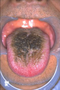
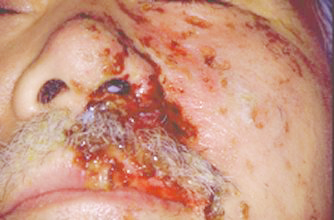
黒毛舌 blackhairytongue.jpg |
カンジダ症 candidiasis.jpg |
頬粘膜血管腫 hemangioma.jpg |
帯状疱疹 herpeszoster.jpg |
帯状疱疹 herpeszoster.jpg |
帯状疱疹 herpeszoster.jpg |
帯状疱疹 herpeszoster.jpg |
1998年 今月の症例
No.1
急性偽膜性カンジダ症
acute pseudomembranous candidiasis
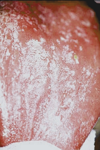
78歳女性、慢性の口内痛とのことで紹介された方です。
受診した翌日に他界されました。
A 78 year-old female presented
to the
Dept. of Dentistry&Oral Surgery with
a three month history of a painful
whole mouth.
She had no smoking history and consumed
no alcohol.
Examination disclosed creamy, pearly white,
or bluish-white patches which can be
scraped off, leaving an erythematous base. She
has
complained of a burning and itching sensation,
and was under treatment with several systemic drugs.
She died of other diseases on the next day of first examination.
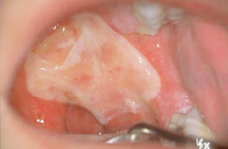
急性偽膜性カンジダ症
Acute pseudomembranous candidiasis
治療:他病死した一例を除いて口腔カンジダ症は
フロリードflorid(持田)ゲル経口用に
ていずれの症例も良好な反応を示し軽快した。
紅斑症
Erythroplakia
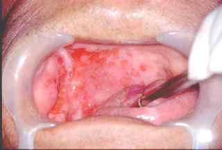
治療:放射線治療および5Fuのいずれの治療にも
良好な反応を示すも患者は口内炎のため治療を拒否、
経過観察中である。
特発性の出血で救急車にて搬送されてきた。
患者は特定の食物(菓子)を摂取すると
発現することを自覚していた。
| 今月の症例 | 資料室 | ニュース | メールサービス | 憩いの部屋 | 歯科リンク
& Top |
歯科リンク
歯科関連の画像の宝庫-(英国) (要登録)
DERWeb
Medlineの検索はこちら-(米国)(無料)
Medline検索
Asahi-net
1998年10月5日初版
ご意見, ご感想はこちらへ
![]()
hf4t-kg@asahi-net.or.jp
更新日2000年10月30日
